PDB 1UBQ · Homo sapiens · Chain A · Model 1
76 modeled residues · 1.80 Å · X-ray diffraction · 8.58 kDa · chain formula mass
Ubiquitin, residues 1–76.
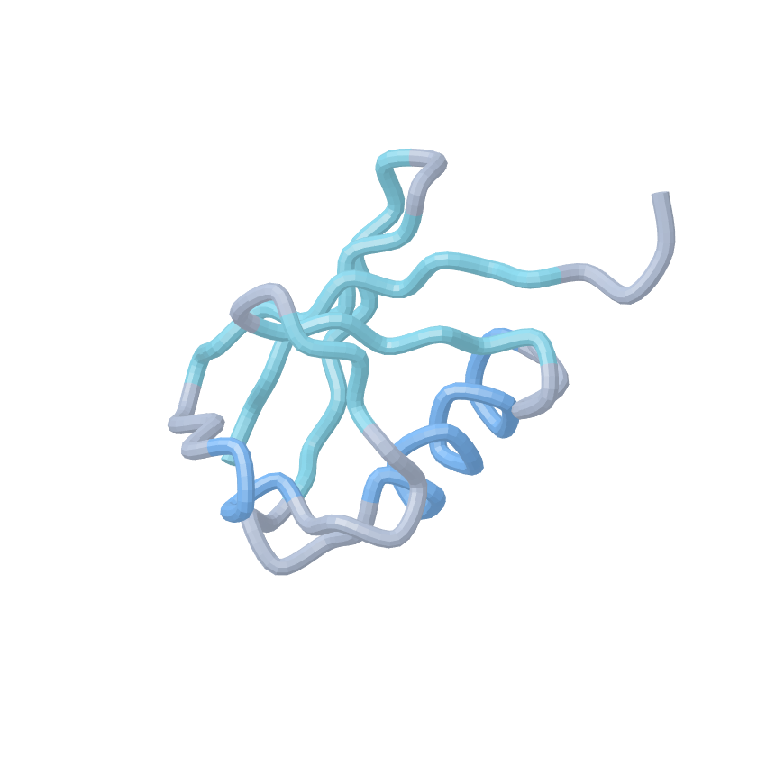
PDB 1A5R · Homo sapiens · Chain A · Model 1
103 modeled residues · Solution NMR · Conformer 1 of 10
GS-extended SUMO-1 precursor, residues −1–101. The precursor includes an N-terminal GS extension and C-terminal tail; it is not mature SUMO-1.
PDB 7PYV · Homo sapiens · Chain C · Model 1
154 modeled residues · 3.27 Å · X-ray diffraction · Two ubiquitin-like domains
FAT10-C0 construct, residues 7–165, from a UBA6 complex. Engineered substitutions: C7T, C9T, C134L, C160S, and C162S. Unresolved residues 82–86 are omitted. The UBA6 partner is not shown.
PDB 2TRX · Escherichia coli · Chain A · Model 1
108 modeled residues · 1.68 Å · X-ray diffraction · Compact α/β fold
Thioredoxin, residues 1–108, with an oxidized Cys32–Cys35 active site. The second crystal copy, copper ions and crystallization additives are not shown.
PDB 4LZT · Gallus gallus · Chain A · Model 1
129 modeled residues · 0.95 Å · X-ray diffraction · Four disulfide bonds in the source
Hen egg-white lysozyme, mature residues 1–129. This backbone view omits disulfide bonds, nitrate ions and solvent.
PDB 1MBN · Physeter macrocephalus · Chain A · Model 1
153 modeled residues · 2.00 Å · X-ray diffraction · Eight α-helices
Sperm-whale myoglobin, residues 1–153. Only the protein backbone is shown; the deposited heme group and hydroxide ligand are omitted.
PDB 1CLL · Homo sapiens · Chain A · Model 1
144 modeled residues · 1.70 Å · X-ray diffraction · Two lobes joined by a central helix
Calcium-bound calmodulin, modeled residues 4–147 of the 148-residue construct. Terminal residues 1–3 and 148 are unresolved; calcium ions and solvent are not shown.